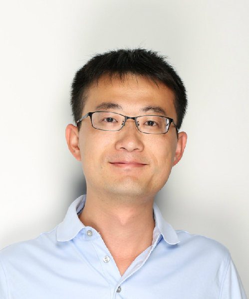
个人介绍:
我的研究主要以电子光学为研究手段,研究基础的细胞生物学问题。
我的科研生涯2009年开始于清华大学结构生物学中心。在此期间主要利用冷冻电镜单颗粒三维重构为技术手段,研究核糖体及其组装机制。通过结构生物学和其他生化手段,获得了核糖体-核糖体组装因子以及一系列不成熟核糖体组装中间体的结构,在此基础上提出了核糖体组装因子的共同特征。
为了拓展电子光学的生物学应用,2014年来到马普生化所Wolfgang Baumeister教授研究组开始博士后训练。主要通过多种不同的技术手段了解神经退行性疾病的细胞毒性来源。 首先,利用单颗粒重构技术,解析了亨廷顿病突变发生蛋白Huntingtin的全长蛋白结构。这是该蛋白被首次鉴定二十五年后第一次获得其结构,对于进一步了解亨廷顿病发病机理有着至关重要的作用。此外,通过优化光镜电镜联用技术和电子断层扫描技术,我们解析了与渐冻症相关的polyGA蛋白聚集物在神经元细胞内的原位结构,并发现该聚集物对蛋白酶体功能的影响,为了解蛋白聚集物的毒性机制提供了新的思路。值得一提的是,通过对整个技术路线的优化,首次实现对蛋白复合物在细胞内原位亚纳米分辨率结构分析。2020年8月正式加入bat365中文官方网站和北大-清华生命科学联合中心,担任研究员,开始组建原位结构生物学实验室。
教育经历:
2009年9月 至2014年7月,博士,清华大学bat365中文官方网站
2005年9月 至2009年7月,本科,兰州大学bat365中文官方网站
工作经历:
2020年8月 至今, 研究员,北大-清华生命科学联合中心
2020年 8月 至今, 研究员,bat365中文官方网站
2014年9月-2020年7月, 博士后, 德国马普生化所(Max-Planck Institute of Biochemistry)
荣誉奖励:
拜耳学者,2021
博雅青年学者,2021
億方学者,2020
Humboldt Fellowship, 2016-2017
EMBO Long-term Fellowship,2015-2017
学术任职:
2021-至今 中国生物物理学会冷冻电镜分会 委员执教课程:
《生命科学与视觉传达》
《三维生物电镜实验技术》
我们是原位结构生物学实验室。关注“细胞建筑学”:各个亚细胞结构是如何搭建成一个具有完整生物学功能的细胞,以及“生物大分子社会学”:细胞内的细胞器、生物大分子之间的相互关系。
原位结构生物学是基于冷冻光电联用(CLEM)、冷冻电子断层扫描(cryo-ET)等技术的新兴结构生物学分支,是一种可以在细胞生理状态下,对生物大分子和亚细胞结构在分子分辨率(1 ~ 10 nm)水平进行原位的结构分析和功能研究的技术手段。我们主要研究方向包括:
• 在纳米、亚纳米尺度对基础细胞生物学问题的研究。
• 对包括神经退行性疾病在内的老龄化疾病致病机制的研究。
• 适用于组织样品的高分辨原位结构生物学方法优化。
* co-first authors, # corresponding author
Wu, Y., Qin, C., Du, W., Guo, Z., Chen, L., Guo, Q.#, 2023. A practical multicellular sample preparation pipeline broadens the application of in situ cryo-electron tomography. Journal of Structural Biology, 107971.
Li, Z., Du, W., Yang, J., Lai, D.-H., Lun, Z.-R., Guo, Q.# (2023). Cryo-Electron Tomography of Toxoplasma gondii Indicates That the Conoid Fiber May Be Derived from Microtubules. Advanced Science 10, 2206595. Cover Story.
Saha, I., Yuste-Checa, P., Da Silva Padilha, M., Guo, Q., Körner, R., Holthusen, H., Trinkaus, V.A., Dudanova, I., Fernández-Busnadiego, R., Baumeister, W., Sanders, D.W., Gautam, S., Diamond, M.I., Hartl, F.U.#, Hipp, M.S.#, (2023). The AAA+ chaperone VCP disaggregates Tau fibrils and generates aggregate seeds in a cellular system. Nature Communications 14, 560.
Li, W.*, Lu, J.*, Xiao, K.*, Zhou, M.*, Li, Y., Zhang, X., Li, Z., Gu, L., Xu, X., Guo, Q.#, Xu, T.#, Ji, W.#, (2023). Integrated multimodality microscope for accurate and efficient target-guided cryo-lamellae preparation. Nature Methods 20, 268-275.
Jiang, W., Wagner, J., Du, W., Plitzko, J., Baumeister, W., Beck, F.#, and Guo, Q.# (2022). A transformation clustering algorithm and its application in polyribosomes structural profiling. Nucleic Acids Research 50, 9001-9011. Cover Story
Riemenschneider, H.*, Guo, Q.*, Bader, J., Frottin, F., Farny, D., Kleinberger, G., Haass, C., Mann, M., Hartl, F.U., Baumeister, W. et al. (2022) Gel-like inclusions of C-terminal fragments of TDP-43 sequester stalled proteasomes in neurons. EMBO Reports, 23, e53890
Peters, J.J., Leitz, J., Guo, Q., Beck, F., Baumeister, W. and Brunger, A.T. (2022) A feature-guided, focused 3D signal permutation method for subtomogram averaging. Journal of Structural Biology, 214, 107851
Huang, B.*, Guo, Q.*, Niedermeier, M.L., Cheng, J., Engler, T., Maurer, M., Pautsch, A., Baumeister, W., Stengel, F., Kochanek, S., et al. (2021). Pathological polyQ expansion does not alter the conformation of the Huntingtin-HAP40 complex. Structure 29, 804-809.e805.
Trinkaus, V.A., Riera-Tur, I., Martínez-Sánchez, A., Bäuerlein, F.J.B., Guo, Q., Arzberger, T., Baumeister, W., Dudanova, I., Hipp, M.S., Hartl, F.U., et al. (2021). In situ architecture of neuronal α-Synuclein inclusions. Nature Communication. 12, 2110.
Seefelder, M., Alva, V., Huang, B., Engler, T., Baumeister, W., Guo, Q., Fernandez-Busnadiego, R., Lupas, A.N., and Kochanek, S. (2020). The evolution of the huntingtin-associated protein 40 (HAP40) in conjunction with huntingtin. BMC Evolutionary Biology 20, 162
Yasuda, S., Tsuchiya, H., Kaiho, A., Guo, Q., Ikeuchi, K., Endo, A., Arai, N., Ohtake, F., Murata, S., Inada, T., et al. (2020). Stress- and ubiquitylation-dependent phase separation of the proteasome. Nature 578, 296–300.
Guo, Q.*, Bin, H.*, Cheng, J., Seefelder, M., Engler, T., Pfeifer, G., Oeckl, P., Otto, M., Moser, F., Maurer, M., Pautsch, A., Baumeister, W.#, Fernandez-Busnadiego, R.#, Kochanek, S.# (2018). The cryo-electron microscopy structure of huntingtin. Nature 555, 117–120.
Guo, Q., Lehmer, C., Martinez-Sanchez, A., Rudack, T., Beck, F., Hartmann, H., Hipp, M.S., Hartl, F.U., Edbauer, D.#, Baumeister, W.#, Fernandez-Busnadiego, R#. (2018) In Situ Structure of Neuronal C9orf72 Poly-GA Aggregates Reveals Proteasome Recruitment. Cell 172, 696-705.e612.
Zhao, Y., Zeng, X., Guo, Q., and Xu, M. (2018). An integration of fast alignment and maximum-likelihood methods for electron subtomogram averaging and classification. Bioinformatics 34, i227-i236.
Li, Z., Guo, Q., Zheng, L., Ji, Y., Xie, Y.T., Lai, D.H., Lun, Z.R., Suo, X., and Gao, N. (2017). Cryo-EM structures of the 80S ribosomes from human parasites Trichomonas vaginalis and Toxoplasma gondii. Cell Research 27, 1275-1288.
Mi, N., Chen, Y., Wang, S., Chen, M., Zhao, M., Yang, G., Ma, M., Su, Q., Luo, S., Shi, J., Xu, J., Guo, Q., et al. (2015). CapZ regulates autophagosomal membrane shaping by promoting actin assembly inside the isolation membrane. Nature Cell Biology 17, 1112-1123.
Feng, B., Mandava, CS., Guo, Q., Wang, J., Cao, W., et al. (2014) Structural and Functional Insights into the Mode of Action of a Universally Conserved Obg GTPase. PLoS Biology 12(5): e1001866.
Yang, Z.*, Guo, Q.*, Goto, S., Chen, Y., Li, N., Muto, A., Himeno, H., Deng, H., Lei, J.#, and Gao, N.# (2014). Characterization of the in vivo 30S ribosomal assembly intermediates reveals essential role of S5 and location of unprocessed ends of the 17S rRNA. Protein Cell 5, 394-407.
Li, N., Chen, Y., Guo, Q., Zhang, Y., Yuan, Y., Ma, C., Deng, H., Lei, J.#, and Gao, N.# (2013). Cryo-EM structures of the late-stage assembly intermediates of the bacterial 50S ribosomal subunit. Nucleic Acids Research 41, 7073-7083.
Guo, Q.*, Goto, S.*, Chen, Y.*, Feng, B., Xu, Y., Muto, A., Himeno, H., Deng, H., Lei, J., and Gao, N. (2013). Dissecting the in vivo assembly of the 30S ribosomal subunit reveals the role of RimM and general features of the assembly process. Nucleic Acids Research 41, 2609-2620.
Huang, W.*, Choi, W.*, Hu, W., Mi, N., Guo, Q., Ma, M., Liu, M., Tian, Y., Lu, P., Wang, F.L., et al. (2012). Crystal structure and biochemical analyses reveal Beclin 1 as a novel membrane binding protein. Cell Research 22, 473-489.
Guo, Q.*, Yuan, Y.*, Xu, Y., Feng, B., Liu, L., Chen, K., Sun, M., Yang, Z., Lei, J.#, and Gao, N.# (2011). Structural basis for the function of a small GTPase RsgA on the 30S ribosomal subunit maturation revealed by cryoelectron microscopy. PNAS 108, 13100-13105.
我们是原位结构生物学实验室。关注“细胞建筑学”:各个亚细胞结构是如何搭建成一个具有完整生物学功能的细胞,以及“生物大分子社会学”:细胞内的细胞器、生物大分子之间的相互关系。
原位结构生物学是基于冷冻光电联用(CLEM)、冷冻电子断层扫描(cryo-ET)等技术的新兴结构生物学分支,是一种可以在细胞生理状态下,对生物大分子和亚细胞结构在分子分辨率(1 ~ 10 nm)水平进行原位的结构分析和功能研究的技术手段。我们主要研究方向包括:
-
在纳米、亚纳米尺度对基础细胞生物学问题的研究。
-
对包括神经退行性疾病在内的老龄化疾病致病机制的研究。
-
病原-宿主相互作用。
-
适用于组织样品的高分辨原位结构生物学方法优化。
 下载Firefox
 下载Firefox

 下载Firefox